Copyright
©The Author(s) 2018.
World J Gastrointest Endosc. Nov 16, 2018; 10(11): 326-339
Published online Nov 16, 2018. doi: 10.4253/wjge.v10.i11.326
Published online Nov 16, 2018. doi: 10.4253/wjge.v10.i11.326
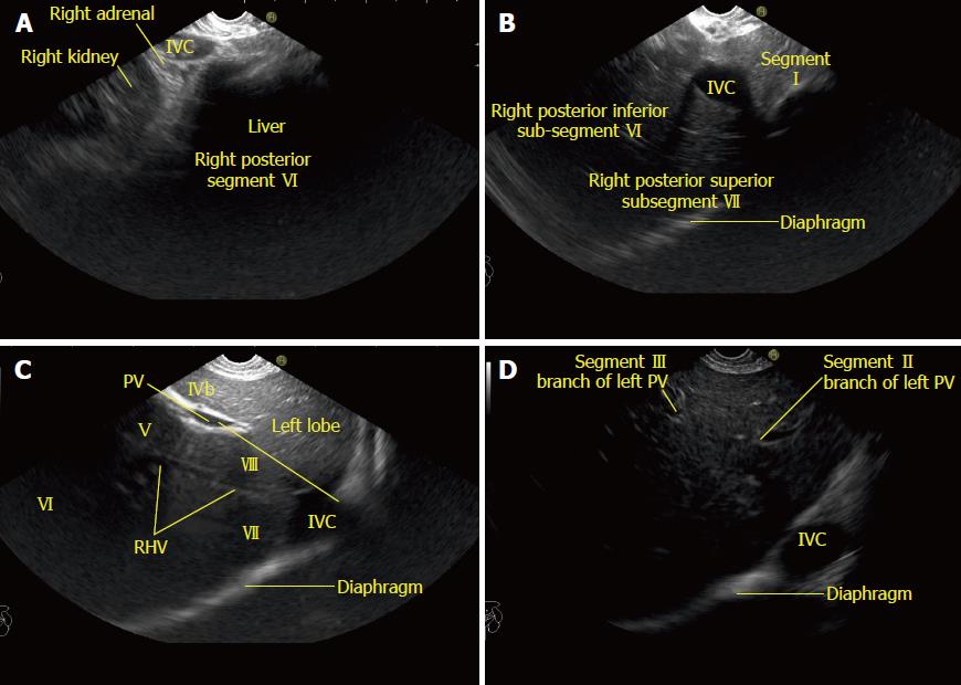
Figure 8 Imaging from the descending duodenum showing the structures visualized during counterclockwise rotation from open position to right.
A: Imaging from the descending duodenum showing the right kidney and inferior vena cava (IVC). The right adrenal gland is seen behind the IVC; B: Imaging from the descending duodenum showing the IVC moving towards the diaphragm. The caudate lobe is seen between the probe and the IVC. The caudate lobe indicates the approximate place of the transverse part of the left branch of the portal vein (PV); C: Imaging from the descending duodenum showing the right hepatic vein. It divides the segments of the right lobe. A line between the cranial end of the IVC and the PV gives approximate locations of the right and left half of the liver; D: The segmental branches (II and III) of the umbilical part of the PV are seen with the IVC at a 4 o’clock position. RHV: Right hepatic vein; IVC: Inferior vena cava; PV: Portal vein.
- Citation: Sharma M, Somani P, Rameshbabu CS, Sunkara T, Rai P. Stepwise evaluation of liver sectors and liver segments by endoscopic ultrasound. World J Gastrointest Endosc 2018; 10(11): 326-339
- URL: https://www.wjgnet.com/1948-5190/full/v10/i11/326.htm
- DOI: https://dx.doi.org/10.4253/wjge.v10.i11.326


