Copyright
©The Author(s) 2018.
World J Gastrointest Endosc. Nov 16, 2018; 10(11): 326-339
Published online Nov 16, 2018. doi: 10.4253/wjge.v10.i11.326
Published online Nov 16, 2018. doi: 10.4253/wjge.v10.i11.326
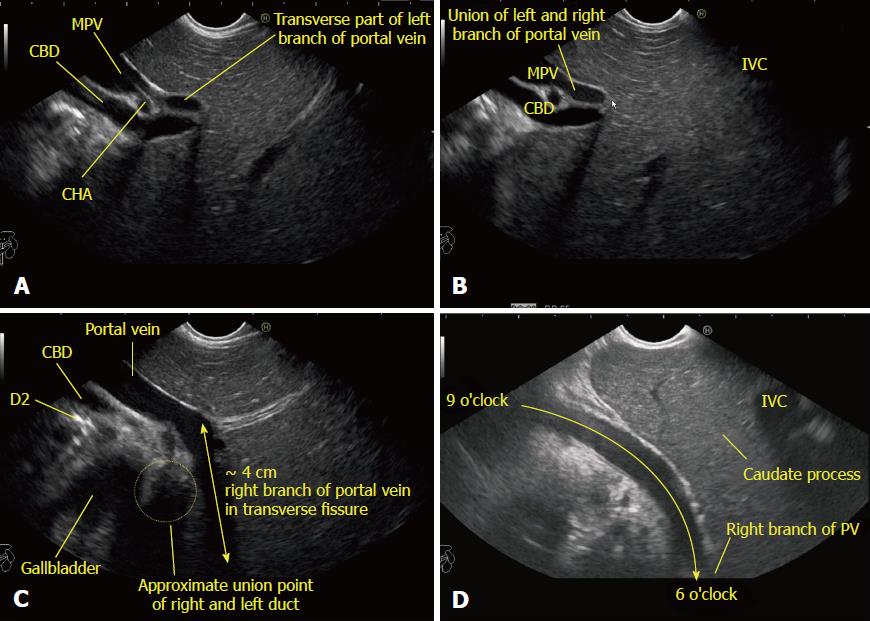
Figure 6 Imaging from neutral position from station 1 showing the tracing of the portal vein and its branches during clockwise rotation from the left to right edge of the transverse fissure.
A: Further clockwise rotation traces the course of the left branch of the portal vein (PV) in the transverse fissure; B: Further rotation shows the union of the right and left branch of the PV; C: The approximate 4 cm breadth of the transverse fissure within which the right branch of the PV joins the left branch; D: The imaging of the right branch of the PV is easy through the caudate process of the liver. With slight up angulation, the right branch of the PV is seen in the transverse fissure going from a 6 o’clock to a 9 o’clock position. CHA: Common hepatic artery; IVC: Inferior vena cava; PV: Portal vein; CBD: Common bile duct; MPV: Main portal vein.
- Citation: Sharma M, Somani P, Rameshbabu CS, Sunkara T, Rai P. Stepwise evaluation of liver sectors and liver segments by endoscopic ultrasound. World J Gastrointest Endosc 2018; 10(11): 326-339
- URL: https://www.wjgnet.com/1948-5190/full/v10/i11/326.htm
- DOI: https://dx.doi.org/10.4253/wjge.v10.i11.326


