Copyright
©The Author(s) 2018.
World J Gastrointest Endosc. Nov 16, 2018; 10(11): 326-339
Published online Nov 16, 2018. doi: 10.4253/wjge.v10.i11.326
Published online Nov 16, 2018. doi: 10.4253/wjge.v10.i11.326
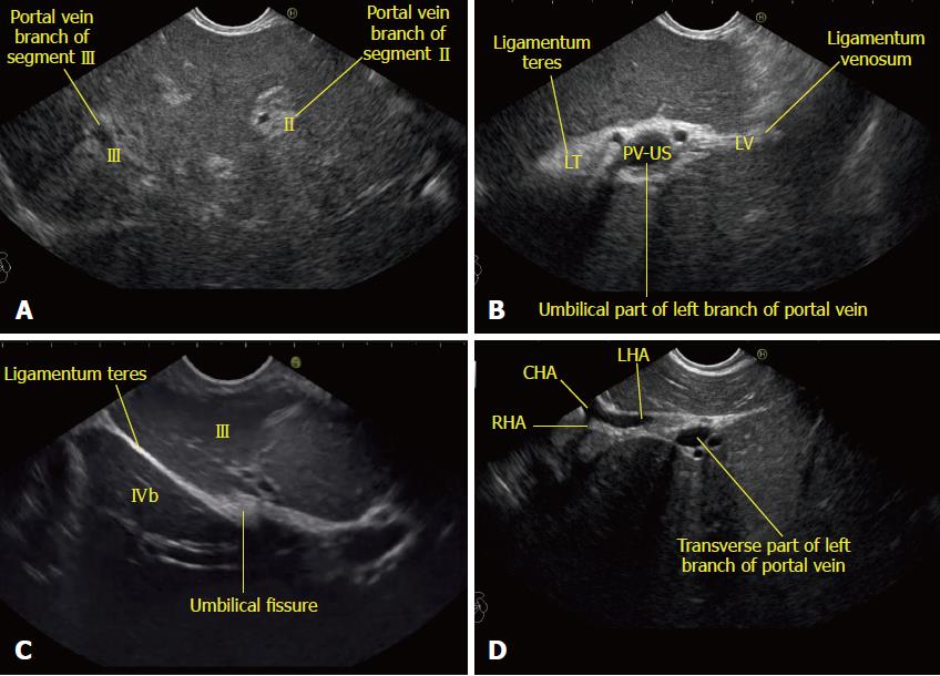
Figure 5 Imaging of liver segments from station 1.
A: Imaging from the abdominal part of esophagus showing segments II and III portal vein branches; B: The fisheye appearance of the umbilical part of the left branch of the portal vein as seen from the abdominal part of esophagus; C: Imaging from the visceral surface of the liver showing that the ligamentum teres is attached to the lower part of the umbilical vein; D: On clockwise rotation near the left edge of the porta hepatis, the umbilical part enters the transverse fissure. At this point the bifurcation of the common hepatic artery can be seen towards the left edge of the transverse fissure. This image shows the entry of the left branch of the common hepatic artery into the transverse fissure. CHA: Common hepatic artery; RHA: Right hepatic artery; LHA: Left hepatic artery.
- Citation: Sharma M, Somani P, Rameshbabu CS, Sunkara T, Rai P. Stepwise evaluation of liver sectors and liver segments by endoscopic ultrasound. World J Gastrointest Endosc 2018; 10(11): 326-339
- URL: https://www.wjgnet.com/1948-5190/full/v10/i11/326.htm
- DOI: https://dx.doi.org/10.4253/wjge.v10.i11.326


