Copyright
©The Author(s) 2018.
World J Gastrointest Endosc. Nov 16, 2018; 10(11): 326-339
Published online Nov 16, 2018. doi: 10.4253/wjge.v10.i11.326
Published online Nov 16, 2018. doi: 10.4253/wjge.v10.i11.326
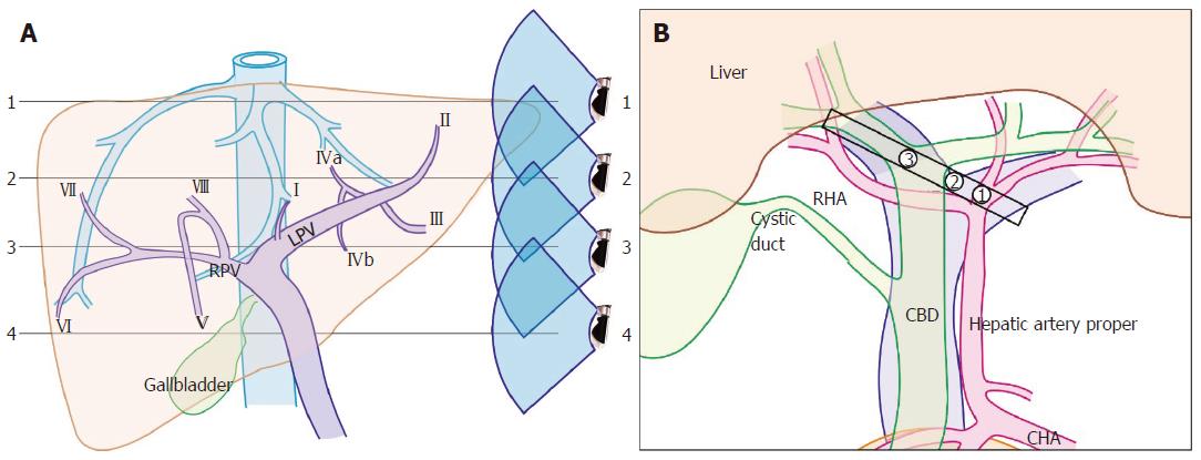
Figure 4 Hepatic vein tributaries with portal vein branches and hilar structures during rotation from left to right edge of transverse fissure.
A: The imaging of hepatic vein tributaries and portal vein branches as the home bases during imaging from the abdominal part of the esophagus and stomach; B: The hilar structures with the divisions during a rotation from the left edge of the transverse fissure to the right edge. (1) hepatic artery into two branches; (2) the union of the right and left branch of the portal vein; (3) the division of the common hepatic duct into right and left branches. LPV: Left branch of the portal vein; RPV: Right branch of the portal vein; CHA: Common hepatic artery; RHA: Right hepatic artery; CBD: Common bile duct.
- Citation: Sharma M, Somani P, Rameshbabu CS, Sunkara T, Rai P. Stepwise evaluation of liver sectors and liver segments by endoscopic ultrasound. World J Gastrointest Endosc 2018; 10(11): 326-339
- URL: https://www.wjgnet.com/1948-5190/full/v10/i11/326.htm
- DOI: https://dx.doi.org/10.4253/wjge.v10.i11.326


