Copyright
©The Author(s) 2018.
World J Gastrointest Endosc. Nov 16, 2018; 10(11): 326-339
Published online Nov 16, 2018. doi: 10.4253/wjge.v10.i11.326
Published online Nov 16, 2018. doi: 10.4253/wjge.v10.i11.326
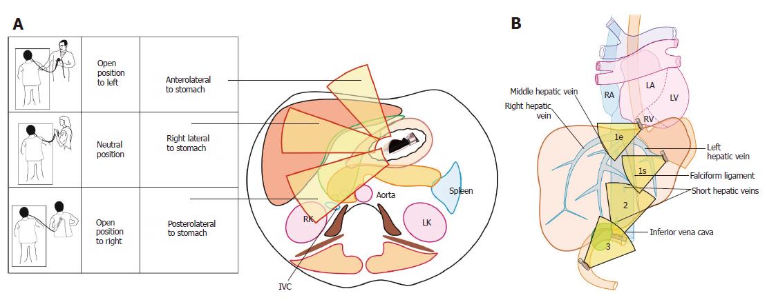
Figure 3 Different operator positions during imaging and three stations of imaging.
A: Rotation is the key movement and should be done in a straight position to transfer the effect of rotation to the tip of the ultrasonic transducer. During imaging from the stomach, the open position to left starts imaging structures placed dorsal to the probe in the esophagus and stomach and primarily screens the left lobe of the liver. The neutral position screens the structures near the liver hilum and the open position to right screens the right lobe of liver; B: The three stations of imaging for liver segments: (1) abdominal part of esophagus (1e) and stomach (1s); (2) duodenal bulb; and (3) second part of the duodenum. IVC: Inferior vena cava; RK: Right kidney; LK: Left kidney; RA: Right atrium; LA: Left atrium; RV: Right ventricle; LV: Left ventricle.
- Citation: Sharma M, Somani P, Rameshbabu CS, Sunkara T, Rai P. Stepwise evaluation of liver sectors and liver segments by endoscopic ultrasound. World J Gastrointest Endosc 2018; 10(11): 326-339
- URL: https://www.wjgnet.com/1948-5190/full/v10/i11/326.htm
- DOI: https://dx.doi.org/10.4253/wjge.v10.i11.326


