Copyright
©The Author(s) 2018.
World J Gastrointest Endosc. Oct 16, 2018; 10(10): 283-293
Published online Oct 16, 2018. doi: 10.4253/wjge.v10.i10.283
Published online Oct 16, 2018. doi: 10.4253/wjge.v10.i10.283
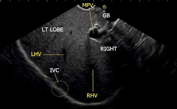
Figure 11 The inferior vena cava is not seen in this image but rotation of the scope shows the approximate area of inferior vena cava (yellow circle) where the left and right hepatic veins merge into inferior vena cava.
MHV: Middle hepatic vein; RHV: Right hepatic vein; LHV: Left hepatic vein; IVC: Inferior vena cava.
- Citation: Sharma M, Somani P, Rameshbabu CS. Linear endoscopic ultrasound evaluation of hepatic veins. World J Gastrointest Endosc 2018; 10(10): 283-293
- URL: https://www.wjgnet.com/1948-5190/full/v10/i10/283.htm
- DOI: https://dx.doi.org/10.4253/wjge.v10.i10.283


