Copyright
©The Author(s) 2018.
World J Gastrointest Endosc. Jan 16, 2018; 10(1): 16-22
Published online Jan 16, 2018. doi: 10.4253/wjge.v10.i1.16
Published online Jan 16, 2018. doi: 10.4253/wjge.v10.i1.16
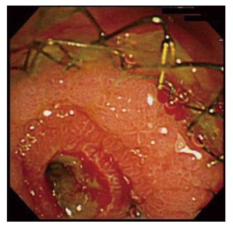
Figure 3 Endoscopic view of the duodenal major papilla after Niti-S 14 placement.
The bile duct cavity is maintained despite bile duct mucosa or tumor growth into the stent.
- Citation: Kikuyama M, Shirane N, Kawaguchi S, Terada S, Mukai T, Sugimoto K. New 14-mm diameter Niti-S biliary uncovered metal stent for unresectable distal biliary malignant obstruction. World J Gastrointest Endosc 2018; 10(1): 16-22
- URL: https://www.wjgnet.com/1948-5190/full/v10/i1/16.htm
- DOI: https://dx.doi.org/10.4253/wjge.v10.i1.16


