Copyright
©The Author(s) 2018.
World J Gastrointest Endosc. Jan 16, 2018; 10(1): 10-15
Published online Jan 16, 2018. doi: 10.4253/wjge.v10.i1.10
Published online Jan 16, 2018. doi: 10.4253/wjge.v10.i1.10
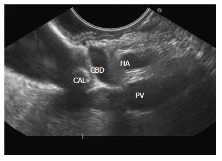
Figure 15 The gall bladder imaging is done from descending duodenum with up deflection and anti-clockwise rotation.
The CBD can be traced and a stone is seen in the Cystic duct. The distended gall bladder is also visualized. PV: Portal vein; CBD: Common bile duct.
- Citation: Sharma M, Somani P, Sunkara T. Imaging of gall bladder by endoscopic ultrasound. World J Gastrointest Endosc 2018; 10(1): 10-15
- URL: https://www.wjgnet.com/1948-5190/full/v10/i1/10.htm
- DOI: https://dx.doi.org/10.4253/wjge.v10.i1.10


