Copyright
©The Author(s) 2018.
World J Gastrointest Endosc. Jan 16, 2018; 10(1): 10-15
Published online Jan 16, 2018. doi: 10.4253/wjge.v10.i1.10
Published online Jan 16, 2018. doi: 10.4253/wjge.v10.i1.10
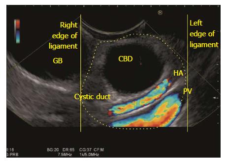
Figure 13 The gall bladder imaging is done from descending duodenum with up deflection and anti-clockwise rotation.
The hepatoduodenal ligament is identified as a bean shaped structure between the probe and liver (shown in dotted yellow area). The CBD can be traced along the cystic duct and the gall bladder which lies outside the right edge of hepatoduodenal ligament. CBD: Common bile duct; GB: Gall bladder.
- Citation: Sharma M, Somani P, Sunkara T. Imaging of gall bladder by endoscopic ultrasound. World J Gastrointest Endosc 2018; 10(1): 10-15
- URL: https://www.wjgnet.com/1948-5190/full/v10/i1/10.htm
- DOI: https://dx.doi.org/10.4253/wjge.v10.i1.10


