Copyright
©The Author(s) 2018.
World J Gastrointest Endosc. Jan 16, 2018; 10(1): 10-15
Published online Jan 16, 2018. doi: 10.4253/wjge.v10.i1.10
Published online Jan 16, 2018. doi: 10.4253/wjge.v10.i1.10
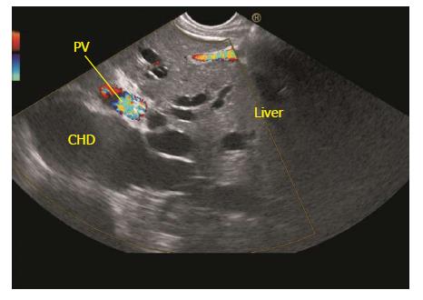
Figure 4 The dilated ducts of segment 2 and 3 can be followed to formation of left hepatic duct.
The left hepatic duct joins the right hepatic duct to form common hepatic duct. The common hepatic duct (CHD) lies beyond the supraduodenal part of portal vein. PV: Portal vein.
- Citation: Sharma M, Somani P, Sunkara T. Imaging of gall bladder by endoscopic ultrasound. World J Gastrointest Endosc 2018; 10(1): 10-15
- URL: https://www.wjgnet.com/1948-5190/full/v10/i1/10.htm
- DOI: https://dx.doi.org/10.4253/wjge.v10.i1.10


