Copyright
©The Author(s) 2018.
World J Gastrointest Endosc. Jan 16, 2018; 10(1): 10-15
Published online Jan 16, 2018. doi: 10.4253/wjge.v10.i1.10
Published online Jan 16, 2018. doi: 10.4253/wjge.v10.i1.10
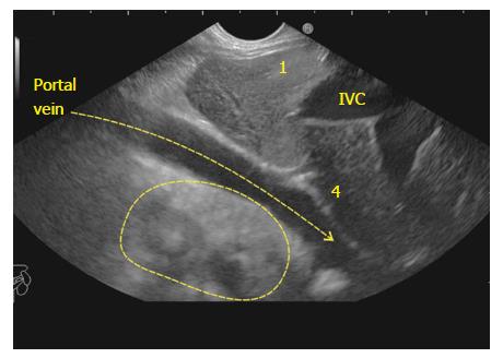
Figure 2 The supraduodenal part of portal vein is seen as a curving vessel going from 5/6 o’clock position to 9/10 o’clock position.
The yellow arrow points to the curving part of portal vein. The area marked with yellow outline shows the area in which the CBD and Gall Bladder can be seen. 1: Segment 1; 4: Segment 4; IVC: Inferior vena cava; CBD: Common bile duct.
- Citation: Sharma M, Somani P, Sunkara T. Imaging of gall bladder by endoscopic ultrasound. World J Gastrointest Endosc 2018; 10(1): 10-15
- URL: https://www.wjgnet.com/1948-5190/full/v10/i1/10.htm
- DOI: https://dx.doi.org/10.4253/wjge.v10.i1.10


