Copyright
©The Author(s) 2018.
World J Gastrointest Endosc. Jan 16, 2018; 10(1): 1-9
Published online Jan 16, 2018. doi: 10.4253/wjge.v10.i1.1
Published online Jan 16, 2018. doi: 10.4253/wjge.v10.i1.1
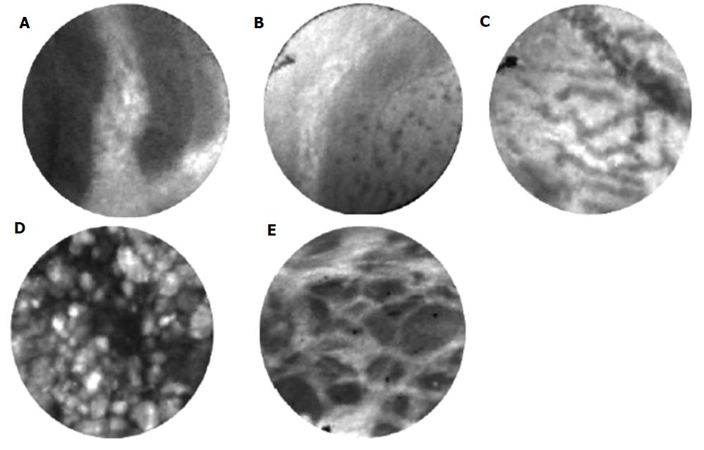
Figure 3 Confocal endomicroscopy findings of various types of pancreatic cystic lesions.
A: Papillae of intraductal papillary mucinous neoplasm; B: Epithelial bands of mucinous cystic neoplasm; C: Fern pattern of serous cystadenoma; D: Bright particles against a dark background of pseudocyst; E: Trabecular pattern of neuroendocrine neoplasm.
- Citation: Li F, Malli A, Cruz-Monserrate Z, Conwell DL, Krishna SG. Confocal endomicroscopy and cyst fluid molecular analysis: Comprehensive evaluation of pancreatic cysts. World J Gastrointest Endosc 2018; 10(1): 1-9
- URL: https://www.wjgnet.com/1948-5190/full/v10/i1/1.htm
- DOI: https://dx.doi.org/10.4253/wjge.v10.i1.1


