Copyright
©The Author(s) 2016.
World J Gastrointest Endosc. Mar 10, 2016; 8(5): 252-258
Published online Mar 10, 2016. doi: 10.4253/wjge.v8.i5.252
Published online Mar 10, 2016. doi: 10.4253/wjge.v8.i5.252
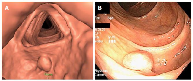
Figure 1 Visualization of a colonic polyp by computed tomographic colonography and optical colonoscopy.
A: Three dimensional view of a splenic flexure colonic polyp on computed tomographic colonography; B: View of the same polyp on optical colonoscopy.
- Citation: El Zoghbi M, Cummings LC. New era of colorectal cancer screening. World J Gastrointest Endosc 2016; 8(5): 252-258
- URL: https://www.wjgnet.com/1948-5190/full/v8/i5/252.htm
- DOI: https://dx.doi.org/10.4253/wjge.v8.i5.252


