Copyright
©The Author(s) 2019.
World J Gastrointest Endosc. Jan 16, 2019; 11(1): 54-60
Published online Jan 16, 2019. doi: 10.4253/wjge.v11.i1.54
Published online Jan 16, 2019. doi: 10.4253/wjge.v11.i1.54
Figure 1 Computed tomography scan revealed a large circumferential mass involving the transverse colon which extended approximately 14 cm in length (arrows).
A: Axial; B: Coronal; C: Transverse.
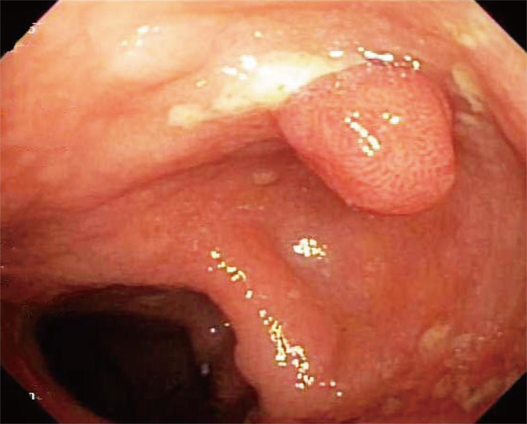
Figure 2 Colonoscopy with multiple polyps and nodularity throughout the colon.
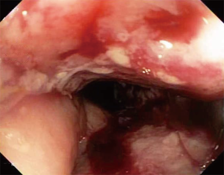
Figure 3 Colonoscopy with 15 cm malignant appearing stricture in the transverse colon.
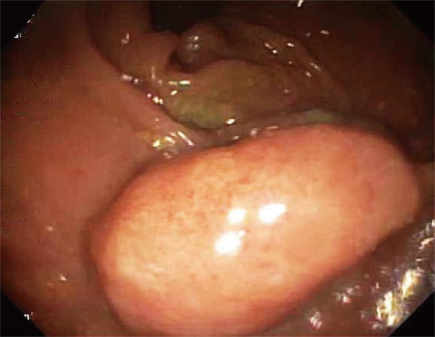
Figure 4 Colonoscopy.
2.5 cm in her rectum polyp.
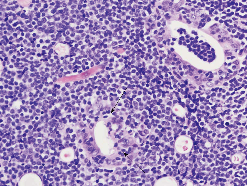
Figure 5 The infiltrate is composed of small to intermediate-sized lymphocytes with round nuclei, inconspicuous nucleoli and lacks an inflammatory background.
Prominent intraepithelial lymphocytosis is present (arrows; Hematoxylin and Eosin, 500 ×). Pathology results of her stomach, duodenum, terminal ileum, colon, transverse stricture and all of her colonic polyps are consistent with enteropathy associated T-cell lymphoma.
- Citation: Fisher A, Yousif E, Piper M. Truth lies below: A case report and literature review of typical appearing polyps yet with an atypical diagnosis. World J Gastrointest Endosc 2019; 11(1): 54-60
- URL: https://www.wjgnet.com/1948-5190/full/v11/i1/54.htm
- DOI: https://dx.doi.org/10.4253/wjge.v11.i1.54


