Copyright
©The Author(s) 2019.
World J Gastrointest Endosc. Jan 16, 2019; 11(1): 22-30
Published online Jan 16, 2019. doi: 10.4253/wjge.v11.i1.22
Published online Jan 16, 2019. doi: 10.4253/wjge.v11.i1.22
Figure 1 Video-cholangiopancreatoscopy shows a normal main pancreatic duct in white light vision and in narrow band imaging mode.
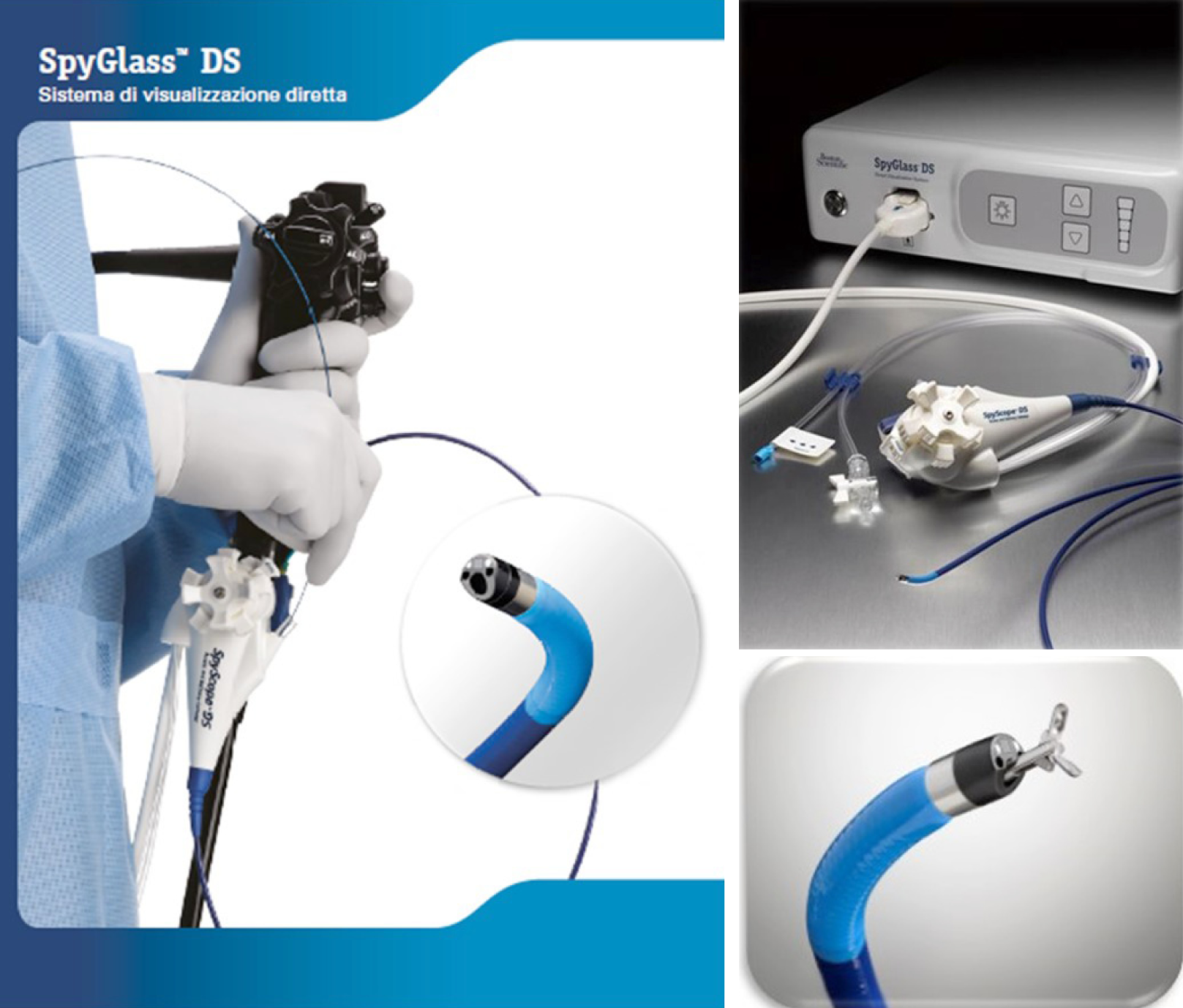
Figure 2 Single-operator video-cholangiopancreatoscopy digital system (SpyGlass DS, Boston Scientific).
Figure 3 Pancreatoscopy with SpyGlass DS.
Figure 4 An ultraslim upper endoscope inserted into the pancreatic duct assisted by a 5-Fr intraductal balloon catheter.
Figure 5 Overtube of a single-balloon enteroscope assisted direct per-oral pancreatoscopy with an ultraslim gastroscope.
Figure 6 Endoscopic classifications of the protruding lesions by per-oral pancreatoscopy.
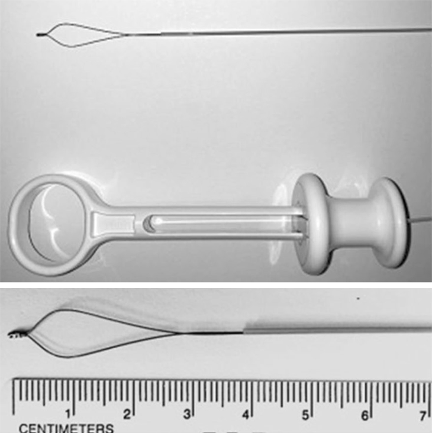
Figure 7 SpyGlassTM Retrieval Snare.
- Citation: De Luca L, Repici A, Koçollari A, Auriemma F, Bianchetti M, Mangiavillano B. Pancreatoscopy: An update. World J Gastrointest Endosc 2019; 11(1): 22-30
- URL: https://www.wjgnet.com/1948-5190/full/v11/i1/22.htm
- DOI: https://dx.doi.org/10.4253/wjge.v11.i1.22


