Copyright
©The Author(s) 2018.
World J Gastrointest Endosc. Sep 16, 2018; 10(9): 145-155
Published online Sep 16, 2018. doi: 10.4253/wjge.v10.i9.145
Published online Sep 16, 2018. doi: 10.4253/wjge.v10.i9.145
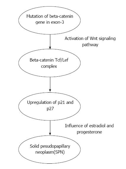
Figure 1 Pathogenesis of solid pseudopapillary neoplasm.
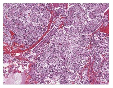
Figure 2 Histological appearance of solid pseudopapillary neoplasm.
A hematoxylin and eosin (H and E) stain of a solid pseudopapillary neoplasm demonstrating eosinophilic neoplastic cells with vacuolated cytoplasm and pseudopapillary appearance.
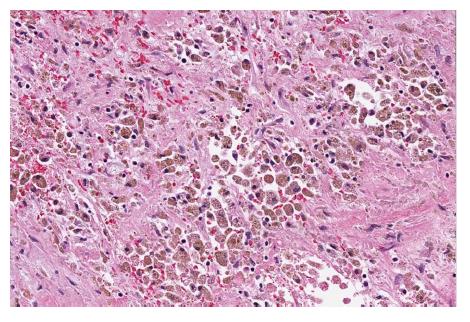
Figure 3 Cytoplasmic and nuclear staining of beta-catenin.
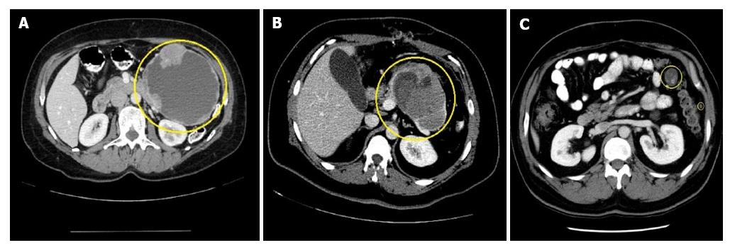
Figure 4 Computed tomography appearances.
A: A pancreatic tail solid pseudopapillary neoplasm. Note the characteristic enhancing solid spaces at the periphery of an encapsulated SPN, accompanied by centrally located cystic space; B: Pancreatic tail solid pseudopapillary neoplasm with septation. The cystic component of SPN with degeneration is characterized by a heterogenous hypoattenuation on CT; C: Abdominal metastatic lesions of SPN. SPN: Solid pseudopapillary neoplasm; CT: Computed tomography.
Figure 5 F-18 fluorodeoxy glucose avid solid pseudopapillary neoplasm metastases and metastatic solid pseudopapillary neoplasm to mesentery.
A: FDG avid SPN metastases to mesentery; B: FDG avid metastatic SPN to mesentery; C: CT appearance of the FDG avid lesion. SPN: Solid pseudopapillary neoplasm; CT: Computed tomography; FDG: F-18 fluorodeoxy glucose.
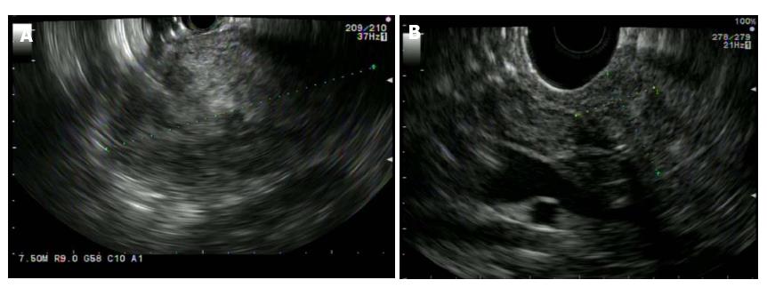
Figure 6 Endoscopic ultrasound appearances.
A: SPN adjacent to the gastric wall. SPN demonstrates heterogenous echogenicity on EUS, with hyperechoic foci representing solid areas with surrounding hypoechoic cystic spaces; B: A pancreatic head SPN. EUS: Endoscopic ultrasound; SPN: Solid pseudopapillary neoplasm.
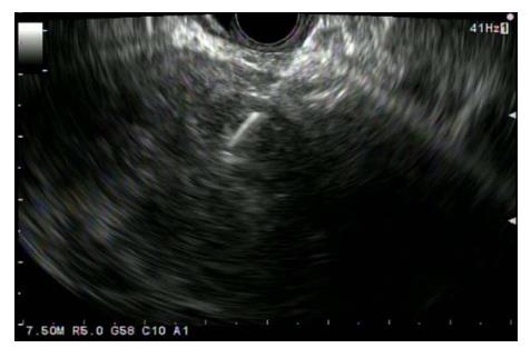
Figure 7 Endoscopic ultrasound guided fine needle aspiration of solid pseudopapillary neoplasm located in body/tail pancreas (Transgastric approach).
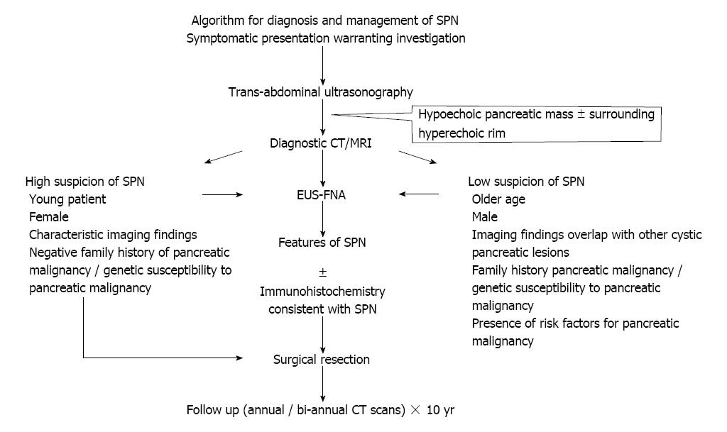
Figure 8 Proposed algorithm for the diagnosis and management of solid pseudopapillary neoplasm.
CT: Computed tomography; MIR: Magnetic resonance imaging; SPN: Solid pseudopapillary neoplasm; EUS-FNA: Endoscopic ultrasound-fine needle aspiration.
- Citation: Lanke G, Ali FS, Lee JH. Clinical update on the management of pseudopapillary tumor of pancreas. World J Gastrointest Endosc 2018; 10(9): 145-155
- URL: https://www.wjgnet.com/1948-5190/full/v10/i9/145.htm
- DOI: https://dx.doi.org/10.4253/wjge.v10.i9.145


