Copyright
©The Author(s) 2017.
World J Hepatol. Dec 28, 2017; 9(36): 1378-1384
Published online Dec 28, 2017. doi: 10.4254/wjh.v9.i36.1378
Published online Dec 28, 2017. doi: 10.4254/wjh.v9.i36.1378
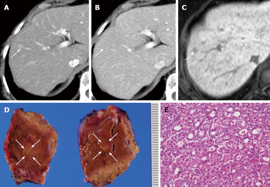
Figure 3 The second tumor.
A: The enhanced tumor of 10 mm in diameter in segment 5 on the early phase in dynamic CT; B: The iso-density tumor on the delayed phase; C: Low intensity tumor on the hepatocyte phase in MRI; D: Cut surface of the 10-mm solid mass in segment 5; E: HE staining showing a pseudoglandular pattern of HCC. CT: Computed tomography; HCC: Hepatocellular carcinoma; HE: Hematoxylin-eosin; MRI: Magnetic resonance imaging.
- Citation: Ide R, Oshita A, Nishisaka T, Nakahara H, Aimitsu S, Itamoto T. Primary biliary cholangitis metachronously complicated with combined hepatocellular carcinoma-cholangiocellular carcinoma and hepatocellular carcinoma. World J Hepatol 2017; 9(36): 1378-1384
- URL: https://www.wjgnet.com/1948-5182/full/v9/i36/1378.htm
- DOI: https://dx.doi.org/10.4254/wjh.v9.i36.1378


