Copyright
©The Author(s) 2017.
World J Hepatol. Dec 8, 2017; 9(34): 1261-1269
Published online Dec 8, 2017. doi: 10.4254/wjh.v9.i34.1261
Published online Dec 8, 2017. doi: 10.4254/wjh.v9.i34.1261
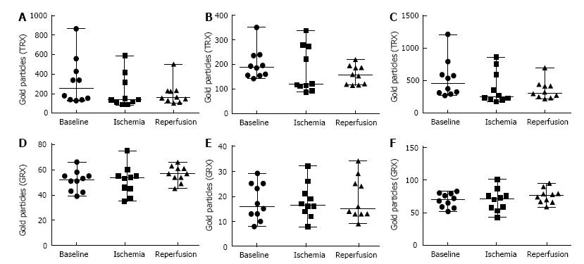
Figure 4 Quantification of immunogold staining.
Number of gold particles of 5 hepatocytes for each time point and patient were recorded. A-C: Values from the TRX immunogold staining where A: Quantification of TRX in the cytosol; B: Quantification of TRX in the nuclei; C: Total value from of TRX both in cytosol and nuclei; D-F: Values from the quantification of GRX immunogold staining where D: Quantification of GRX in the cytosol; E: Quantification of GRX in the nuclei; F: Total value of GRX both in cytosol and nuclei. Baseline: Before induction of ischemia; Ischemia: Twenty minute of ischemia; Reperfusion: Twenty minute after reperfusion.
- Citation: Jawad R, D’souza M, Selenius LA, Lundgren MW, Danielsson O, Nowak G, Björnstedt M, Isaksson B. Morphological alterations and redox changes associated with hepatic warm ischemia-reperfusion injury. World J Hepatol 2017; 9(34): 1261-1269
- URL: https://www.wjgnet.com/1948-5182/full/v9/i34/1261.htm
- DOI: https://dx.doi.org/10.4254/wjh.v9.i34.1261


