Copyright
©The Author(s) 2017.
World J Hepatol. Dec 8, 2017; 9(34): 1261-1269
Published online Dec 8, 2017. doi: 10.4254/wjh.v9.i34.1261
Published online Dec 8, 2017. doi: 10.4254/wjh.v9.i34.1261
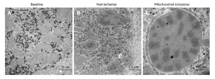
Figure 2 Crystalline mitochondrial inclusions.
A: Baseline, before induction of ischemia, shows hepatocyte mitochondria with normal appearance; B: Post-ischemia, showing mitochondria with the crystalline inclusions and a few dilated mitochondria; C: Mitochondrial inclusions, close-up of a single mega-mitochondrion showing the inclusions post-ischemia.
- Citation: Jawad R, D’souza M, Selenius LA, Lundgren MW, Danielsson O, Nowak G, Björnstedt M, Isaksson B. Morphological alterations and redox changes associated with hepatic warm ischemia-reperfusion injury. World J Hepatol 2017; 9(34): 1261-1269
- URL: https://www.wjgnet.com/1948-5182/full/v9/i34/1261.htm
- DOI: https://dx.doi.org/10.4254/wjh.v9.i34.1261


