Copyright
©The Author(s) 2017.
World J Hepatol. Dec 8, 2017; 9(34): 1261-1269
Published online Dec 8, 2017. doi: 10.4254/wjh.v9.i34.1261
Published online Dec 8, 2017. doi: 10.4254/wjh.v9.i34.1261
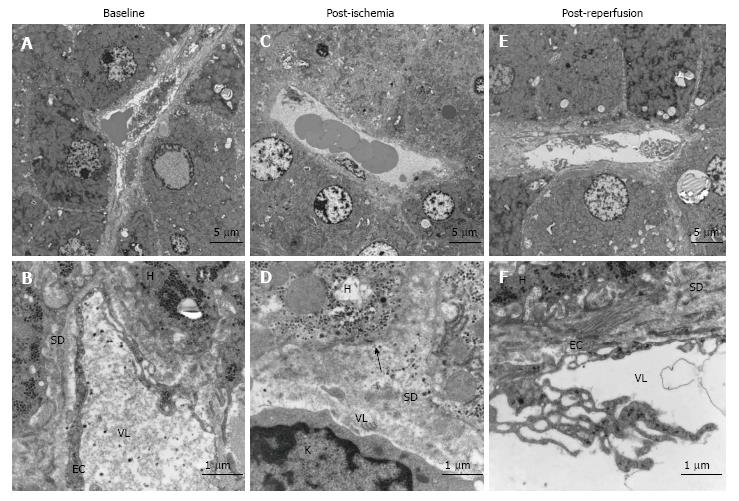
Figure 1 Morphological changes in liver before and after ischemia and reperfusion, transmission electron micrographs of representative images of liver sections from one patient.
A: Baseline, before induction of ischemia, shows the normal state of liver morphology at a cellular level; B: Morphology of a sinusoid with neighbouring hepatocytes 20 min after ischemia; C: Representative image of sinusoid and haptocytes 20 min after reperfusion; D: Endothelial lining of a sinusoid with hepatocyte microvilli; E: Morphology of the Space of Disse post-ischemia; F: Morphology of the space of disse post-reperfusion. SD: Space of disse; H: Hepatocyte; EC: Endothelial cells; VL: Vessel lumen; K: Kupffer cell. Arrow shows the absence of hepatocyte microvilli.
- Citation: Jawad R, D’souza M, Selenius LA, Lundgren MW, Danielsson O, Nowak G, Björnstedt M, Isaksson B. Morphological alterations and redox changes associated with hepatic warm ischemia-reperfusion injury. World J Hepatol 2017; 9(34): 1261-1269
- URL: https://www.wjgnet.com/1948-5182/full/v9/i34/1261.htm
- DOI: https://dx.doi.org/10.4254/wjh.v9.i34.1261


