Copyright
©The Author(s) 2017.
World J Hepatol. Nov 18, 2017; 9(32): 1227-1238
Published online Nov 18, 2017. doi: 10.4254/wjh.v9.i32.1227
Published online Nov 18, 2017. doi: 10.4254/wjh.v9.i32.1227
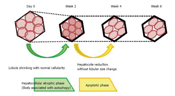
Figure 7 Schema of the histological changes occurring following interruption of portal blood flow.
At 2 wk after obstruction of the portal vein, lobular shrinkage was observed without reduction in hepatocyte number, but with strong LC3 and LAMP2 expression. These changes may be associated with autophagy, and this process is termed the hepatocellular atrophic phase. At week 4, hepatocyte numbers fell, without a reduction in lobule size, but with an elevation of TUNEL staining. These secondary changes may be attributed to apoptosis occurring after autophagy, characterizing the hepatocellular atrophic phase. No significant histological changes were observed at week 6 compared with week 4.
- Citation: Iwao Y, Ojima H, Kobayashi T, Kishi Y, Nara S, Esaki M, Shimada K, Hiraoka N, Tanabe M, Kanai Y. Liver atrophy after percutaneous transhepatic portal embolization occurs in two histological phases: Hepatocellular atrophy followed by apoptosis. World J Hepatol 2017; 9(32): 1227-1238
- URL: https://www.wjgnet.com/1948-5182/full/v9/i32/1227.htm
- DOI: https://dx.doi.org/10.4254/wjh.v9.i32.1227


