Copyright
©The Author(s) 2017.
World J Hepatol. Nov 18, 2017; 9(32): 1227-1238
Published online Nov 18, 2017. doi: 10.4254/wjh.v9.i32.1227
Published online Nov 18, 2017. doi: 10.4254/wjh.v9.i32.1227
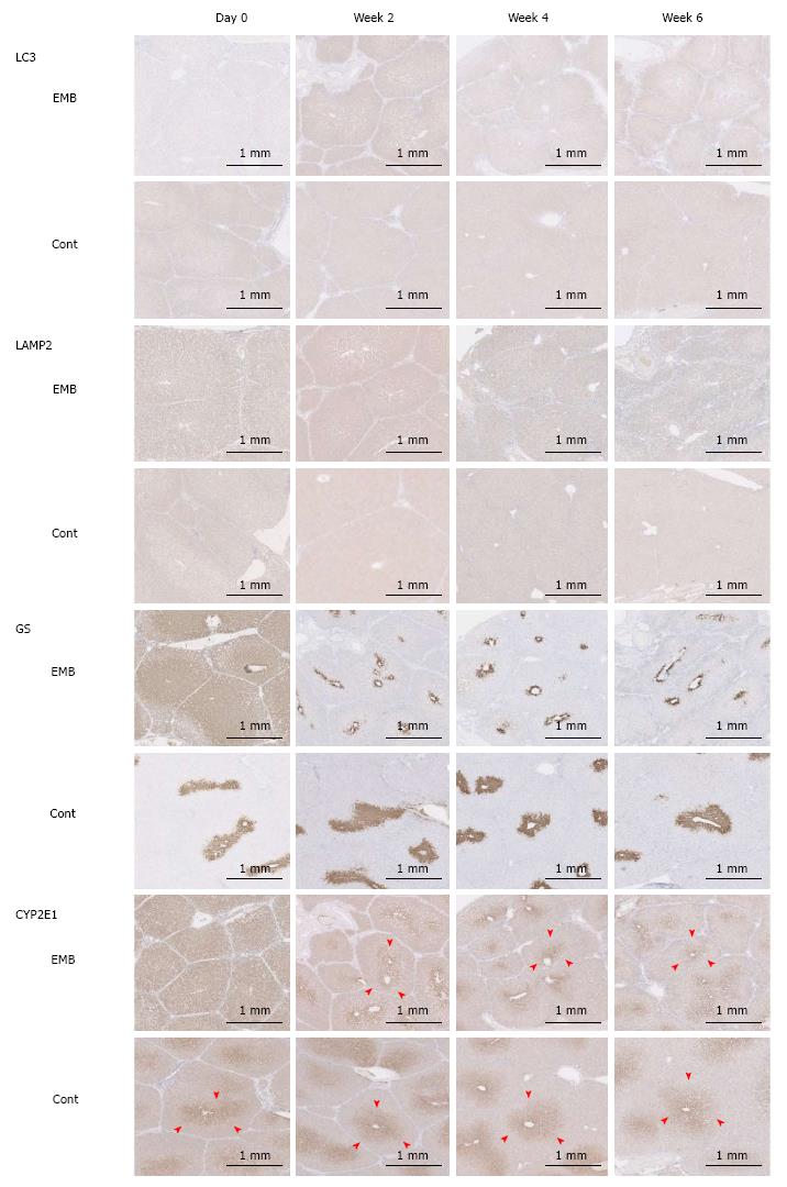
Figure 4 Light chain 3, lysosomal-associated membrane protein 2, glutamine synthetase, and cytochrome P450 2E1 immunohistochemical staining intensities.
Expression of LC3 and LAMP2 was highest in the embolized segment of porcine specimens at 2 wk after PTPE. Zonation of GS and CYP2E1 (arrowheads) was expanded in the embolized area immediately after interruption of portal blood flow, but was reduced in the embolized lobe at 2 wk. EMB: Embolized area; Cont: Control lobe area; LC3: Light chain 3; LAMP2: Lysosomal-associated membrane protein 2; GS: Glutamine synthetase; CYP2E1: Cytochrome P450 2E1; IHC: Immunohistochemical.
- Citation: Iwao Y, Ojima H, Kobayashi T, Kishi Y, Nara S, Esaki M, Shimada K, Hiraoka N, Tanabe M, Kanai Y. Liver atrophy after percutaneous transhepatic portal embolization occurs in two histological phases: Hepatocellular atrophy followed by apoptosis. World J Hepatol 2017; 9(32): 1227-1238
- URL: https://www.wjgnet.com/1948-5182/full/v9/i32/1227.htm
- DOI: https://dx.doi.org/10.4254/wjh.v9.i32.1227


