Copyright
©The Author(s) 2017.
World J Hepatol. Nov 18, 2017; 9(32): 1227-1238
Published online Nov 18, 2017. doi: 10.4254/wjh.v9.i32.1227
Published online Nov 18, 2017. doi: 10.4254/wjh.v9.i32.1227
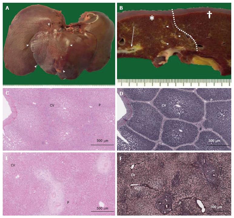
Figure 2 Macroscopic images of pig livers following interruption of portal blood flow and structural comparison of pig and human liver lobules.
A: Liver removed from a pig at week 2 after PTPE. Segmental macroscopic atrophy could be seen (arrowheads); B: Portal thrombosis was observed at the cut surface (arrow) in the embolized area (*), and fibrous thickening of the portal areas was observed (arrowheads) compared with nonembolized area (†). A clear border (dotted line) was observed between these areas; C-F: Microscopic views (C, D: Pig; E, F: Human) show central veins (CV) and portal areas (P) in HE-stained sections (C, E), but these features are more clearly observed in silver-stained sections (D, F). Lobule structure was less well defined in human specimens (F).
- Citation: Iwao Y, Ojima H, Kobayashi T, Kishi Y, Nara S, Esaki M, Shimada K, Hiraoka N, Tanabe M, Kanai Y. Liver atrophy after percutaneous transhepatic portal embolization occurs in two histological phases: Hepatocellular atrophy followed by apoptosis. World J Hepatol 2017; 9(32): 1227-1238
- URL: https://www.wjgnet.com/1948-5182/full/v9/i32/1227.htm
- DOI: https://dx.doi.org/10.4254/wjh.v9.i32.1227


