Copyright
©The Author(s) 2017.
World J Hepatol. Sep 18, 2017; 9(26): 1081-1091
Published online Sep 18, 2017. doi: 10.4254/wjh.v9.i26.1081
Published online Sep 18, 2017. doi: 10.4254/wjh.v9.i26.1081
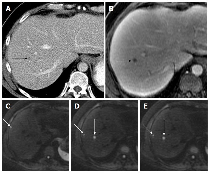
Figure 1 Value of diffusion-weighted magnetic resonance imaging in lesion detection in a 51-year-old male with metastatic leiomyosarcoma of the thigh.
A: Axial contrast enhanced CT scan demonstrated a subtle hypodensity in the right lobe of liver (black arrow); B: Axial post gadolinium T1-weighted MR image demonstrates a single metastatic lesion (black arrow); C-E: DW-MR image at b-600 demonstrates additional lesions (white arrows). DW-MR: Diffusion-weighted magnetic resonance; CT: Computed tomography.
- Citation: Shenoy-Bhangle A, Baliyan V, Kordbacheh H, Guimaraes AR, Kambadakone A. Diffusion weighted magnetic resonance imaging of liver: Principles, clinical applications and recent updates. World J Hepatol 2017; 9(26): 1081-1091
- URL: https://www.wjgnet.com/1948-5182/full/v9/i26/1081.htm
- DOI: https://dx.doi.org/10.4254/wjh.v9.i26.1081


