Copyright
©The Author(s) 2017.
World J Hepatol. Jul 8, 2017; 9(19): 840-849
Published online Jul 8, 2017. doi: 10.4254/wjh.v9.i19.840
Published online Jul 8, 2017. doi: 10.4254/wjh.v9.i19.840
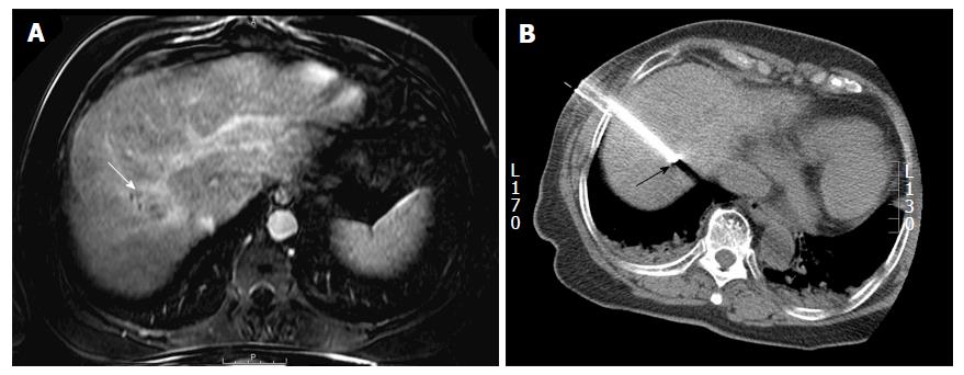
Figure 4 Percutaneous radiofrequency ablation of a hepatic dome hepatocellular carcinoma in a 54-year-old man.
A: Axial post gadolinium T1-weighted image in the portal venous phase demonstrates a 3.4 cm hepatocellular carcinoma in the hepatic dome (arrow); B: During the radiofrequency ablation procedure, the patient was placed in the oblique position and using a lateral intercostal approach the tumor was accessed for a successful ablation (arrow).
- Citation: Kambadakone A, Baliyan V, Kordbacheh H, Uppot RN, Thabet A, Gervais DA, Arellano RS. Imaging guided percutaneous interventions in hepatic dome lesions: Tips and tricks. World J Hepatol 2017; 9(19): 840-849
- URL: https://www.wjgnet.com/1948-5182/full/v9/i19/840.htm
- DOI: https://dx.doi.org/10.4254/wjh.v9.i19.840


