Copyright
©The Author(s) 2017.
World J Hepatol. Jun 8, 2017; 9(16): 733-745
Published online Jun 8, 2017. doi: 10.4254/wjh.v9.i16.733
Published online Jun 8, 2017. doi: 10.4254/wjh.v9.i16.733
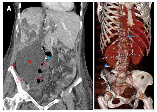
Figure 6 Biloma in a 49-year-old female patient who underwent associating liver partition and portal vein ligation for staged hepatectomy because of peripheral cholangiocarcinoma of the right liver lobe.
A: Computed tomography was performed because of bile flowing from the right drainage (blue triangle). The examination confirmed a large fluid collection beneath the DH (red triangle), which distended the plastic bag (red arrows). Biloma was removed with the DH during stage 2 surgery, resolving the biliary leakage originating from right transection surface; B: Normal position of the two drains on volume rendering reconstruction. Left drain has a vertical course along the line of transection up to the inferior margin of the diaphragm (arrow). Right drain has an horizontal course beneath DH (triangle), with its his placed within the plastic bag, in order to drain collections. DH: Diseased hemiliver.
- Citation: Zerial M, Lorenzin D, Risaliti A, Zuiani C, Girometti R. Abdominal cross-sectional imaging of the associating liver partition and portal vein ligation for staged hepatectomy procedure. World J Hepatol 2017; 9(16): 733-745
- URL: https://www.wjgnet.com/1948-5182/full/v9/i16/733.htm
- DOI: https://dx.doi.org/10.4254/wjh.v9.i16.733


