Copyright
©The Author(s) 2017.
World J Hepatol. Jun 8, 2017; 9(16): 733-745
Published online Jun 8, 2017. doi: 10.4254/wjh.v9.i16.733
Published online Jun 8, 2017. doi: 10.4254/wjh.v9.i16.733
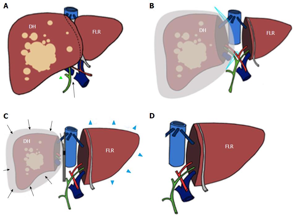
Figure 1 Scheme of trisectionectomy associating liver partition and portal vein ligation procedure.
During surgical stage 1 the right portal vein is sectioned and sutured (arrow in A) after performing cholecystectomy (green triangle in A). Subsequently, the diseased hemiliver (DH) is sectioned from the future liver remnant (FLR) and wrapped with a bag (B). At the time of surgical stage 2 (C), hypertrophy of the (FLR) (blue arrowheads in D) and atrophy of the DH (arrows) have been obtained. Associating liver partition and portal vein ligation for staged hepatectomy procedure is then completed by removing the DH (D).
- Citation: Zerial M, Lorenzin D, Risaliti A, Zuiani C, Girometti R. Abdominal cross-sectional imaging of the associating liver partition and portal vein ligation for staged hepatectomy procedure. World J Hepatol 2017; 9(16): 733-745
- URL: https://www.wjgnet.com/1948-5182/full/v9/i16/733.htm
- DOI: https://dx.doi.org/10.4254/wjh.v9.i16.733


