Copyright
©The Author(s) 2016.
World J Hepatol. Mar 18, 2016; 8(8): 411-420
Published online Mar 18, 2016. doi: 10.4254/wjh.v8.i8.411
Published online Mar 18, 2016. doi: 10.4254/wjh.v8.i8.411
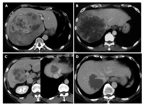
Figure 1 Abdominal computed tomography scan images of cases 1-4.
A: Case 1: Lumen of the IVC compressed by a huge right liver tumor with expansive growth; B: Case 2: A large right liver tumor involving the right aspect of the IVC and the confluence of middle and left hepatic veins; C: Case 3: Two tumors were present, a right liver tumor involving the right portal vein and the portal vein bifurcation, and another tumor occupying the cranial part of segments 4 and 8 and involving the left aspect of the trunk of the middle and left hepatic veins; D: Case 4: A tumor involving the right dorsal aspect of the IVC and its confluence of the right hepatic vein. IVC: Inferior vena cava.
- Citation: Ko S, Kirihataya Y, Matsumoto Y, Takagi T, Matsusaka M, Mukogawa T, Ishikawa H, Watanabe A. Retrocaval liver lifting maneuver and modifications of total hepatic vascular exclusion for liver tumor resection. World J Hepatol 2016; 8(8): 411-420
- URL: https://www.wjgnet.com/1948-5182/full/v8/i8/411.htm
- DOI: https://dx.doi.org/10.4254/wjh.v8.i8.411


