Copyright
©The Author(s) 2016.
World J Hepatol. Feb 8, 2016; 8(4): 231-262
Published online Feb 8, 2016. doi: 10.4254/wjh.v8.i4.231
Published online Feb 8, 2016. doi: 10.4254/wjh.v8.i4.231
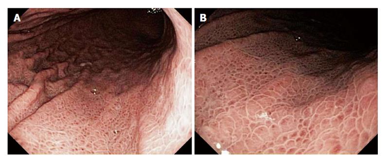
Figure 2 A 60-year-old man presented for routine endoscopic screening for esophageal varices due to a history of Child-Pugh class B cirrhosis, with a model for end-stage liver disease score = 18, from hepatitis C secondary to former intravenous drug use.
The patient denied a history of gastrointestinal bleeding. The hematocrit was 40.1%. Esophagogastroduodenoscopy revealed the classic findings of portal hypertensive gastropathy, including a pale white reticular (mosaic) pattern surrounding small polygonal areas of mucosa, with variable erythema, in the entire stomach, but most prominently in the gastric fundus and body. B is a relatively close-up view of the lesions seen in A.
- Citation: Gjeorgjievski M, Cappell MS. Portal hypertensive gastropathy: A systematic review of the pathophysiology, clinical presentation, natural history and therapy. World J Hepatol 2016; 8(4): 231-262
- URL: https://www.wjgnet.com/1948-5182/full/v8/i4/231.htm
- DOI: https://dx.doi.org/10.4254/wjh.v8.i4.231


