Copyright
©The Author(s) 2016.
World J Hepatol. Oct 18, 2016; 8(29): 1222-1233
Published online Oct 18, 2016. doi: 10.4254/wjh.v8.i29.1222
Published online Oct 18, 2016. doi: 10.4254/wjh.v8.i29.1222
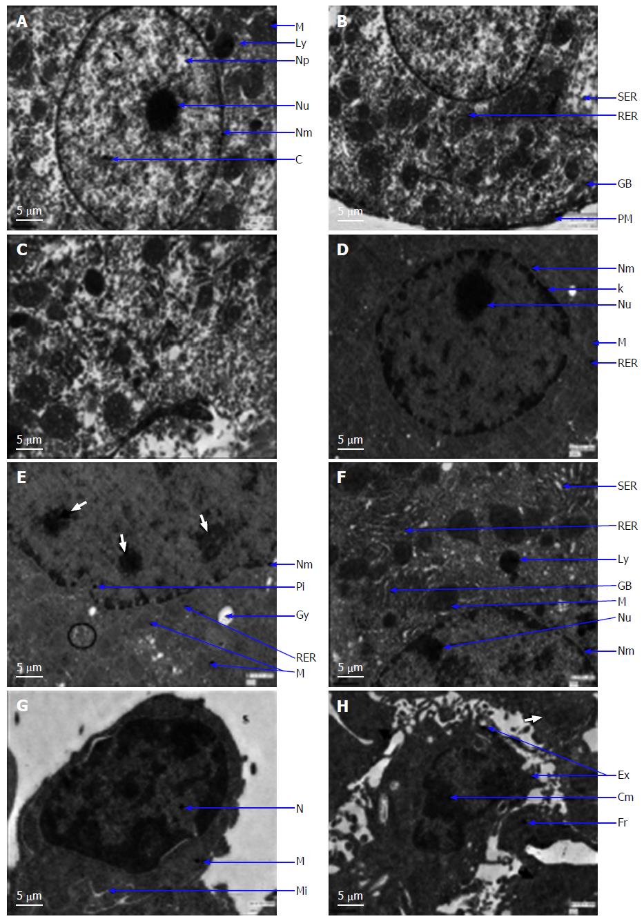
Figure 2 Transmission electron micrographs (× 2550) of liver tissue.
A and B: Control group illustrating round nucleus (N), nuclear membrane (Nm), nucleolus (Nu), and chromatin (C) and nucleoplasm (Np). Clear cytoplasm with mitochondria (M), dark bodies as lysosomes (Ly), smooth endoplasmic reticulum (SER), rough endoplasmic reticulum (RER), and Ggolgi bodies (GB) surrounded by plasma membrane (PM) were observed; C: LycT group illustrating similar characteristics to those in theof control liver micrograph; D and E: NDEA group illustrating deformed N with disrupted and convoluted Nm, darkly masses in the nucleolus (white arrows), deposition of karyotin (k) and pseudo-inclusions (Pi). Cytoplasm was also found to be compactly packed with multiple pleomorphic M and RER. Percentage of other organelles was found to be lower, and variable light and dark granules of fat (encircled) and glycogen (Gy) deposits were also observed; F-H: LycT + NDEA group illustrating perforated nuclear membrane. Cytoplasm containedwas occupied with pleomorphic mitochondria, with ahowever higher percentage of organelles such as GB, Ly, RER and higher glycogen deposits. Macrophages in the sinusoid (S) were characterized by a large nucleus to cytoplasmic ratio, abundant mitochondria and microvilli (Mi). Apoptotic body characterized by cell shrinkage and condensation of nuclear chromatin into delineated masses (Cm), forming extensions (Ex), and crowded with closely packed cellular organelles (white arrow) and fragments of nucleus (Fr). LycT: Lycopene extracted from tomatoes; NDEA: N-Nitrosodiethylamine.
- Citation: Gupta P, Bhatia N, Bansal MP, Koul A. Lycopene modulates cellular proliferation, glycolysis and hepatic ultrastructure during hepatocellular carcinoma. World J Hepatol 2016; 8(29): 1222-1233
- URL: https://www.wjgnet.com/1948-5182/full/v8/i29/1222.htm
- DOI: https://dx.doi.org/10.4254/wjh.v8.i29.1222


