Copyright
©The Author(s) 2016.
World J Hepatol. Oct 8, 2016; 8(28): 1157-1168
Published online Oct 8, 2016. doi: 10.4254/wjh.v8.i28.1157
Published online Oct 8, 2016. doi: 10.4254/wjh.v8.i28.1157
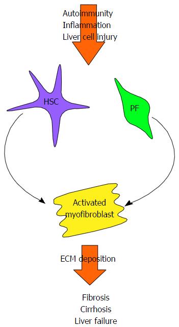
Figure 2 Development of fibrosis and cirrhosis in autoimmune liver disease.
Persistent autoimmune-mediated inflammation and liver cell injury leads to the activation and differentiation of quiescent hepatic stellate cells (HSC) and portal fibroblasts (PF) into activated myofibroblasts. These proliferative, pro-inflammatory and pro-fibrogenic myofibroblasts increase collagen synthesis and deposit extracellular matrix proteins (ECM), leading to the development of fibrous scar tissue. Cirrhosis, characterised by significant fibrosis and nodular regeneration, eventually ensues, with the resultant loss of liver function and eventually liver failure.
- Citation: Liberal R, Grant CR. Cirrhosis and autoimmune liver disease: Current understanding. World J Hepatol 2016; 8(28): 1157-1168
- URL: https://www.wjgnet.com/1948-5182/full/v8/i28/1157.htm
- DOI: https://dx.doi.org/10.4254/wjh.v8.i28.1157


