Copyright
©The Author(s) 2016.
World J Hepatol. Sep 8, 2016; 8(25): 1047-1060
Published online Sep 8, 2016. doi: 10.4254/wjh.v8.i25.1047
Published online Sep 8, 2016. doi: 10.4254/wjh.v8.i25.1047
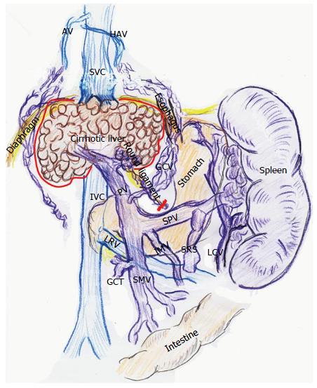
Figure 1 Vascular alterations in advanced liver cirrhosis.
Collaterals along the round ligament are removed with native liver (red line). Collaterals developed around the native liver are also ligated (red line). AV: Azygos vein; GCT: Gastro-colic trunk; GCV: Gastric coronary vein; HAV: Hemi-azygos vein; IMV: Inferior mesenteric vein; IVC: Inferior vena cava; LCV: Left colic vein; LRV: Left renal vein; PV: Portal vein; SMV: Superior mesenteric vein; SPV: Splenic vein; SRS: Splenorenal shunt; SVC: Superior vena cava.
- Citation: Hori T, Ogura Y, Onishi Y, Kamei H, Kurata N, Kainuma M, Takahashi H, Suzuki S, Ichikawa T, Mizuno S, Aoyama T, Ishida Y, Hirai T, Hayashi T, Hasegawa K, Takeichi H, Ota A, Kodera Y, Sugimoto H, Iida T, Yagi S, Taniguchi K, Uemoto S. Systemic hemodynamics in advanced cirrhosis: Concerns during perioperative period of liver transplantation. World J Hepatol 2016; 8(25): 1047-1060
- URL: https://www.wjgnet.com/1948-5182/full/v8/i25/1047.htm
- DOI: https://dx.doi.org/10.4254/wjh.v8.i25.1047


