Copyright
©The Author(s) 2016.
World J Hepatol. Jul 8, 2016; 8(19): 796-814
Published online Jul 8, 2016. doi: 10.4254/wjh.v8.i19.796
Published online Jul 8, 2016. doi: 10.4254/wjh.v8.i19.796
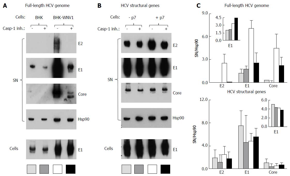
Figure 13 Caspase-1 inhibitor conditionally inhibits the secretion of hepatitis C virus particles by baby hamster kidney-West Nile virus cells.
A: BHK-21 and BHK-WNV cells were transfected with a mix of HCVbp-coding and P2B plasmids; the next day, a caspase-1 inhibitor was added in the culture medium and the cells were incubated for 2 more days; cell lysates and HCV particles were harvested and analyzed by Western blot (WB); B: BHK-WNV cells were transfected with a plasmid coding for the structural (core, E1, E2) genes of HCV H77 strain, plus (+) or minus (-) p7, then were analyzed as in (A); C: Quantification of WBs: Top panels for (A) and bottom panels for (B); bar inside patterns are displayed underneath corresponding data; error bars represent standard deviations; inserts = results in cells. HCV: Hepatitis C virus; BHK-WNV: Baby hamster kidney-West Nile virus.
- Citation: Triyatni M, Berger EA, Saunier B. Assembly and release of infectious hepatitis C virus involving unusual organization of the secretory pathway. World J Hepatol 2016; 8(19): 796-814
- URL: https://www.wjgnet.com/1948-5182/full/v8/i19/796.htm
- DOI: https://dx.doi.org/10.4254/wjh.v8.i19.796


