Copyright
©The Author(s) 2016.
World J Hepatol. Jul 8, 2016; 8(19): 796-814
Published online Jul 8, 2016. doi: 10.4254/wjh.v8.i19.796
Published online Jul 8, 2016. doi: 10.4254/wjh.v8.i19.796
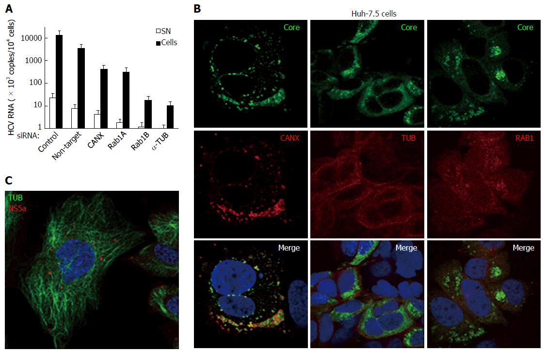
Figure 12 Hepatitis C virus produced in cultured human hepatic cells involves same cellular factors as those enhancing hepatitis C virus production in baby hamster kidney-West Nile virus cells.
A: Huh-7.5 cells were seeded on 24-well plates and the next day were transfected, or not, with siRNA, as indicated. After 2 d, cells were reseeded into 24-well plates and, the next day, incubated at a MOI = 0.5 with HCVcc produced with the JFH-1 strain in Huh-7.5 cells. At 3 dpi, HCV RNA contents were determined in the cells (closed bars) and SN (open bars) by RT-qPCR (TaqMan). Results are plotted on a log-scale; errors bars represent maximum variations observed in this assay; B: Huh-7.5 cells were electroporated with in vitro-transcribed genome of the JFH-1 strain and passaged for two weeks, then were seeded onto coverslips. Two days later, IF was performed with anti-HCV core 7-50 (green) and anti-human CANX, tubulin (TUB) or RAB1 antibodies (red); C: Huh-7.5 cells were inoculated with HCVbp-4cys produced in permissive BHK-WNV cells (8); 2 d later, the cells were incubated with both ReASH (red) and Taxol fluorophore conjugate (green); result representative of two independent experiments; B and C: Nuclei were counterstained with DAPI and cells were observed by confocal microscopy. CANX: Calnexin; HCV: Hepatitis C virus; BHK-WNV: Baby hamster kidney-West Nile virus; SN: Supernatants.
- Citation: Triyatni M, Berger EA, Saunier B. Assembly and release of infectious hepatitis C virus involving unusual organization of the secretory pathway. World J Hepatol 2016; 8(19): 796-814
- URL: https://www.wjgnet.com/1948-5182/full/v8/i19/796.htm
- DOI: https://dx.doi.org/10.4254/wjh.v8.i19.796


