Copyright
©The Author(s) 2016.
World J Hepatol. Jul 8, 2016; 8(19): 796-814
Published online Jul 8, 2016. doi: 10.4254/wjh.v8.i19.796
Published online Jul 8, 2016. doi: 10.4254/wjh.v8.i19.796
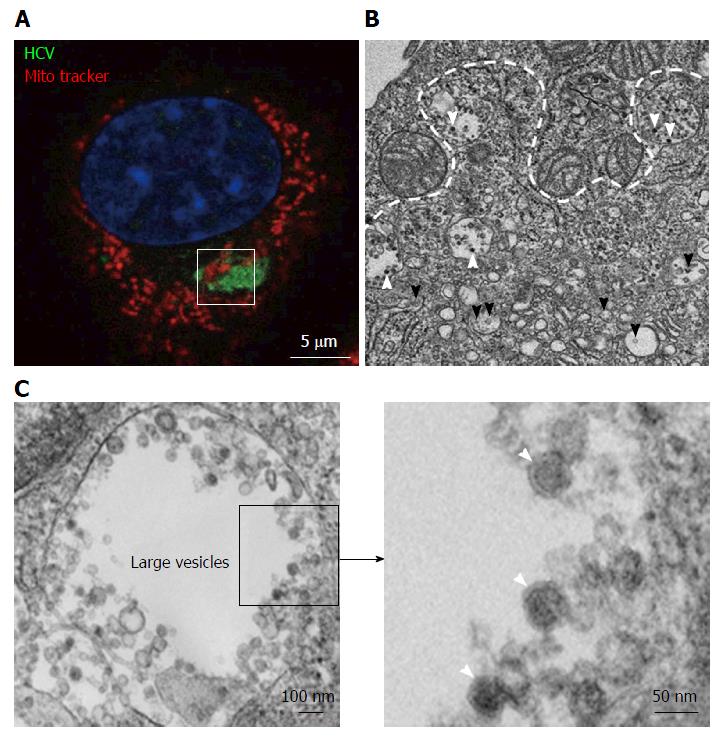
Figure 2 Transmission electron microscopy view of a hepatitis C virus compartment forming in baby hamster kidney-West Nile virus cells cells.
BHK-WNV cells were transfected with a mix of HCVbp-expressing and P2B plasmids (8). A: IF analysis showing a compartment recognized by human serum HCV IgG (green) surrounded by mitochondria (red); nucleus is counterstained with DAPI (blue). The white square delimits an area similar to that displayed in the next panel; B: Thin section observed by transmission electron microscopy: White arrowheads: Electron-dense viral particles in large vesicles; black arrowheads: Nascent viral particles in traffic vesicles; white dotted line: Limit of mitochondria surrounding the compartment containing viral particles (cf. area within white square in previous panel); C: Left panel: Example of large vesicle that develops in the cytoplasm of permissive BHK-WNV cells upon expression of full-length HCV genome; Right panel: Magnification of viral particles. HCV: Hepatitis C virus; BHK-WNV: Baby hamster kidney-West Nile virus.
- Citation: Triyatni M, Berger EA, Saunier B. Assembly and release of infectious hepatitis C virus involving unusual organization of the secretory pathway. World J Hepatol 2016; 8(19): 796-814
- URL: https://www.wjgnet.com/1948-5182/full/v8/i19/796.htm
- DOI: https://dx.doi.org/10.4254/wjh.v8.i19.796


