Copyright
©The Author(s) 2016.
World J Hepatol. Jun 28, 2016; 8(18): 779-784
Published online Jun 28, 2016. doi: 10.4254/wjh.v8.i18.779
Published online Jun 28, 2016. doi: 10.4254/wjh.v8.i18.779
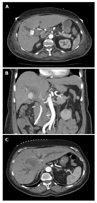
Figure 1 Computerized tomography scan demonstrating pseudoaneurysm and significant biliary ductal dilatation.
A: Triple contrast computerized tomography scan of abdomen demonstrates hepatic artery pseuodoaneurysm; B: With significant thrombus formation adjacent to aneurysm; C: Significant biliary ductal dilatation in the right and left hepatic ducts.
- Citation: Luckhurst CM, Perez C, Collinsworth AL, Trevino JG. Atypical presentation of a hepatic artery pseudoaneurysm: A case report and review of the literature. World J Hepatol 2016; 8(18): 779-784
- URL: https://www.wjgnet.com/1948-5182/full/v8/i18/779.htm
- DOI: https://dx.doi.org/10.4254/wjh.v8.i18.779


