Copyright
©The Author(s) 2016.
World J Hepatol. Jun 18, 2016; 8(17): 731-738
Published online Jun 18, 2016. doi: 10.4254/wjh.v8.i17.731
Published online Jun 18, 2016. doi: 10.4254/wjh.v8.i17.731
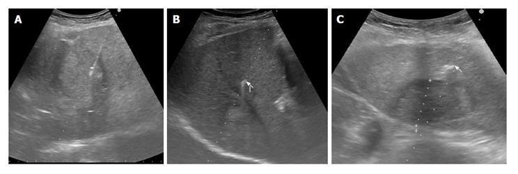
Figure 2 Ultrasonography-guided fiducial placement.
The two different gold markers are sonographically undistinguishable. A: Needle delivering fiducial into a liver mass; B: Hyperechoic flexible wire notched gold marker, 0.28 mm × 10 mm, Gold Anchor marker (arrow) near a liver mass; C: Hyperechoic Grain cylindrical gold marker, 1 mm × 4 mm (arrow) near a liver mass.
- Citation: Marsico M, Gabbani T, Livi L, Biagini MR, Galli A. Therapeutic usability of two different fiducial gold markers for robotic stereotactic radiosurgery of liver malignancies: A pilot study. World J Hepatol 2016; 8(17): 731-738
- URL: https://www.wjgnet.com/1948-5182/full/v8/i17/731.htm
- DOI: https://dx.doi.org/10.4254/wjh.v8.i17.731


