Copyright
©The Author(s) 2015.
World J Hepatol. May 8, 2015; 7(7): 926-941
Published online May 8, 2015. doi: 10.4254/wjh.v7.i7.926
Published online May 8, 2015. doi: 10.4254/wjh.v7.i7.926
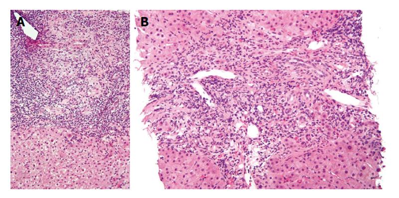
Figure 1 Lower (A) and higher (B) magnification photomicrographs of a liver biopsy sample in a patient with early primary biliary cirrhosis shows a moderately severe, mixed inflammatory infiltrate consisting mostly of lymphocytes and plasma cells concentrated around small bile ducts in the portal area.
Note the absence of inflammation around hepatocytes (H and E stains). Both figures obtained with permission (granted under the GNU Free Documentation License, Version 1.2) from: http://commons.wikimedia.org/wiki/File:Primary_biliary_cirrhosis_intermed_mag_2.jpg. Accessed March 10, 2015.
- Citation: Purohit T, Cappell MS. Primary biliary cirrhosis: Pathophysiology, clinical presentation and therapy. World J Hepatol 2015; 7(7): 926-941
- URL: https://www.wjgnet.com/1948-5182/full/v7/i7/926.htm
- DOI: https://dx.doi.org/10.4254/wjh.v7.i7.926


