Copyright
©The Author(s) 2015.
World J Hepatol. Apr 28, 2015; 7(6): 874-884
Published online Apr 28, 2015. doi: 10.4254/wjh.v7.i6.874
Published online Apr 28, 2015. doi: 10.4254/wjh.v7.i6.874
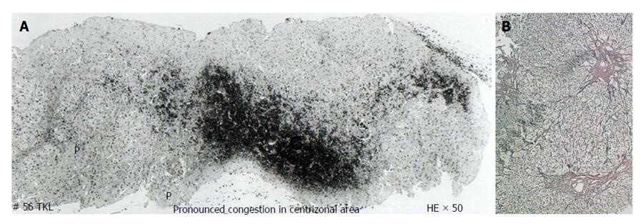
Figure 8 Histology showing fibrosis in centri-lobular areas.
A: Histology of liver of a patient with hepatic vena cava syndrome during acute exacerbation showing acute congestive changes around central vein (c) and sparing of liver around portal tract (P) due to hepatic venous outflow obstruction; B: Histology of liver of patient with hepatic vena cava syndrome a few months after development of hepatic venous outflow obstruction during acute exacerbation showing fibrosis around central vein. Histology of liver of a patient with hepatic vena cava syndrome showing the wall of a thrombosed medium sized intra-hepatic vein that occurred during acute exacerbation. HE: Hematoxylin eosin stain.
- Citation: Shrestha SM. Liver cirrhosis in hepatic vena cava syndrome (or membranous obstruction of inferior vena cava). World J Hepatol 2015; 7(6): 874-884
- URL: https://www.wjgnet.com/1948-5182/full/v7/i6/874.htm
- DOI: https://dx.doi.org/10.4254/wjh.v7.i6.874


