Copyright
©The Author(s) 2015.
World J Hepatol. Apr 28, 2015; 7(6): 874-884
Published online Apr 28, 2015. doi: 10.4254/wjh.v7.i6.874
Published online Apr 28, 2015. doi: 10.4254/wjh.v7.i6.874
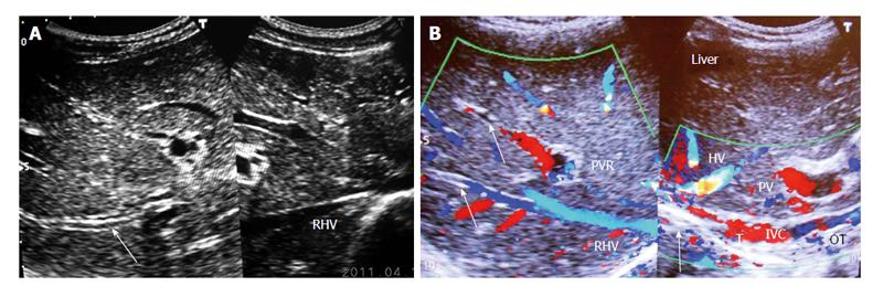
Figure 3 Ultrasonography showing thrombosed intra-hepatic veins.
A: Ultrasonography showing diffuse thrombosed and echoic walls of large and medium-sized intra-hepatic veins (one of which is indicated by an arrow) that occurred during acute exacerbation. Right hepatic vein (RHV) orifice is narrowed; B: Color Doppler Ultrasonography of patient with cirrhosis. It shows long segment stenosis of inferior vena cava (IVC) with recent thrombus (T) and old organized thrombi (OT) on thick posterior wall. Arrow shows thrombosed large and medium-sized intra-hepatic veins. HV: Hepatic vein; PV: Portal vein; PVR: Portal vein radical.
- Citation: Shrestha SM. Liver cirrhosis in hepatic vena cava syndrome (or membranous obstruction of inferior vena cava). World J Hepatol 2015; 7(6): 874-884
- URL: https://www.wjgnet.com/1948-5182/full/v7/i6/874.htm
- DOI: https://dx.doi.org/10.4254/wjh.v7.i6.874


