Copyright
©The Author(s) 2015.
World J Hepatol. Apr 28, 2015; 7(6): 874-884
Published online Apr 28, 2015. doi: 10.4254/wjh.v7.i6.874
Published online Apr 28, 2015. doi: 10.4254/wjh.v7.i6.874
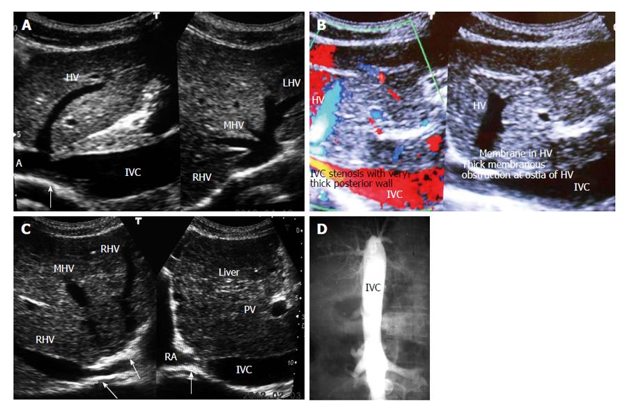
Figure 1 Inferior vena cava obstruction.
A: Ultrasonography showing stenosis of inferior vena cava (IVC) at cavo-atrial junction. Note patent orifices hepatic vein (HV)-right HV (RHV), middle HV (MHV) and left HV (LHV); B: Color Doppler ultrasonography of a patient with liver cirrhosis showing IVC stenosis, membranes in HV; C: Ultrasonography of a patient with liver cirrhosis showing complete obstruction of IVC at cavo-atrial junction and obstruction at orifices of MHV and LHV. Note dilated hepatic veins; D: Cavogram showing complete obstruction of the IVC. PV: Portal vein; RA: Right atrium.
- Citation: Shrestha SM. Liver cirrhosis in hepatic vena cava syndrome (or membranous obstruction of inferior vena cava). World J Hepatol 2015; 7(6): 874-884
- URL: https://www.wjgnet.com/1948-5182/full/v7/i6/874.htm
- DOI: https://dx.doi.org/10.4254/wjh.v7.i6.874


