Copyright
©The Author(s) 2015.
World J Hepatol. Mar 27, 2015; 7(3): 468-487
Published online Mar 27, 2015. doi: 10.4254/wjh.v7.i3.468
Published online Mar 27, 2015. doi: 10.4254/wjh.v7.i3.468
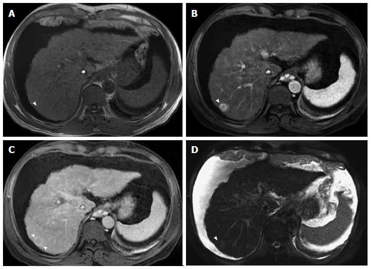
Figure 16 Ring-enhancing hepatocellular carcinoma, indicative of a more aggressive course.
A: In-phase GRE T1 weighted image; B: Post-contrast fat-suppressed 3D-GRE T1-weighted images during the (B) late hepatic arterial and (C) delayed phases; (D) Fat-suppressed SSFSE T2-weighted image. There is a small nodule at hepatic segment #7, which demonstrates iso to slightly low T1 signal (arrowhead, A), heterogeneous increased arterial enhancement, predominantly peripheral (arrowhead, B), washout and pseudocapsule enhancement on delayed images (arrowhead, C), and mildly increased T2 signal intensity (arrowhead, D) in keeping with ring-enhancing HCC. HCC: Hepatocellular carcinoma; GRE: Gradient recalled echo; SSFSE: Single-shot fast spin-echo.
- Citation: Watanabe A, Ramalho M, AlObaidy M, Kim HJ, Velloni FG, Semelka RC. Magnetic resonance imaging of the cirrhotic liver: An update. World J Hepatol 2015; 7(3): 468-487
- URL: https://www.wjgnet.com/1948-5182/full/v7/i3/468.htm
- DOI: https://dx.doi.org/10.4254/wjh.v7.i3.468


