Copyright
©The Author(s) 2015.
World J Hepatol. Mar 27, 2015; 7(3): 468-487
Published online Mar 27, 2015. doi: 10.4254/wjh.v7.i3.468
Published online Mar 27, 2015. doi: 10.4254/wjh.v7.i3.468
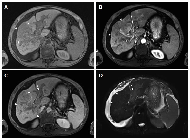
Figure 15 Diffuse hepatocellular carcinoma associated with tumor thrombus.
(A) Pre- and post-contrast fat-suppressed 3D-GRE T1-weighted images during the (B) late hepatic arterial and (C) delayed phases; (D) Fat-suppressed SSFSE T2-weighted image. There is a large ill-defined area involving the right hepatic lobe, which shows decreased T1 signal in pre-contrast images (arrowheads, A), heterogeneous mildly increased arterial enhancement (arrowheads, B), and heterogeneous delayed washout with permeative appearance (arrowheads, C), and heterogeneous mildly increased T2 signal (arrowhead, D) in keeping with HCC. There is also evidence of expansion of the portal vein branches, abnormal increased arterial enhancement (arrow, B), lack of opacification and washout on the delayed images (arrow, C), and mildly increased T2 signal (arrow, D) simulating the behavior of the primary tumor in keeping with diffuse tumor thrombus, a finding almost invariably associated with diffuse HCC subtype. Also of note is the mild to moderate ascites and omental hypertrophy secondary to portal hypertension. HCC: Hepatocellular carcinoma; GRE: Gradient recalled echo.
- Citation: Watanabe A, Ramalho M, AlObaidy M, Kim HJ, Velloni FG, Semelka RC. Magnetic resonance imaging of the cirrhotic liver: An update. World J Hepatol 2015; 7(3): 468-487
- URL: https://www.wjgnet.com/1948-5182/full/v7/i3/468.htm
- DOI: https://dx.doi.org/10.4254/wjh.v7.i3.468


