Copyright
©The Author(s) 2015.
World J Hepatol. Mar 27, 2015; 7(3): 468-487
Published online Mar 27, 2015. doi: 10.4254/wjh.v7.i3.468
Published online Mar 27, 2015. doi: 10.4254/wjh.v7.i3.468
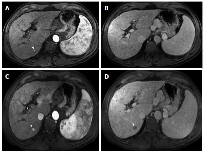
Figure 9 Dysplastic nodule progressing into an hepatocellular carcinoma.
Post-contrast fat-suppressed 3D-GRE T1-weighted images during the late hepatic arterial (A and C) and delayed phases (B and D). There is a small right hepatic lobe nodule, which demonstrates increased arterial enhancement (arrowhead, A) and fades out on the delayed images (B) on the initial examination. On the 4-month follow-up study, there is evidence of interval growth (arrowhead, C, D) and development of clear delayed washout (arrowhead, D) both of which are signs of progression into HCC. HCC: Hepatocellular carcinoma; GRE: Gradient recalled echo.
- Citation: Watanabe A, Ramalho M, AlObaidy M, Kim HJ, Velloni FG, Semelka RC. Magnetic resonance imaging of the cirrhotic liver: An update. World J Hepatol 2015; 7(3): 468-487
- URL: https://www.wjgnet.com/1948-5182/full/v7/i3/468.htm
- DOI: https://dx.doi.org/10.4254/wjh.v7.i3.468


