Copyright
©The Author(s) 2015.
World J Hepatol. Mar 27, 2015; 7(3): 468-487
Published online Mar 27, 2015. doi: 10.4254/wjh.v7.i3.468
Published online Mar 27, 2015. doi: 10.4254/wjh.v7.i3.468
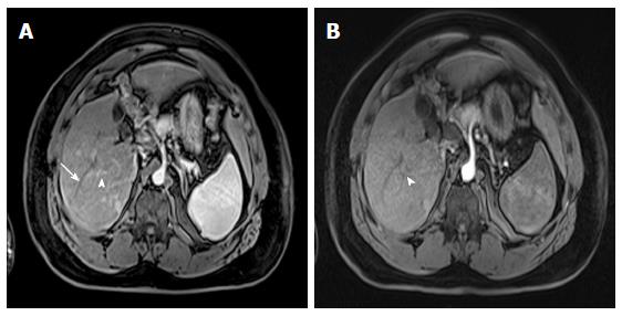
Figure 2 Value of proper timing for detecting hypervascular hepatic lesions.
A-B: Post-contrast fat-suppressed 3D-GRE T1-weighted images acquired 4 mo apart. A: Initial scanning shows contrast in the portal vain branches (arrowhead, A), without opacification of the hepatic veins (arrow, A), suggesting late hepatic arterial phase timing; the optimal time for detecting hypervascular pathologies, with demonstration of multiple lesions; B: A subsequent scan acquired 4 mo later shows contrast in the hepatic artery without opacification of the portal vein branches (arrowhead, B), suggesting an early arterial timing, without evidence of hypervascular lesions. A subsequent scan was acquired (not shown); which confirmed the persistence of these hypervascular lesions. GRE: Gradient recalled echo.
- Citation: Watanabe A, Ramalho M, AlObaidy M, Kim HJ, Velloni FG, Semelka RC. Magnetic resonance imaging of the cirrhotic liver: An update. World J Hepatol 2015; 7(3): 468-487
- URL: https://www.wjgnet.com/1948-5182/full/v7/i3/468.htm
- DOI: https://dx.doi.org/10.4254/wjh.v7.i3.468


