Copyright
©The Author(s) 2015.
World J Hepatol. Nov 18, 2015; 7(26): 2664-2675
Published online Nov 18, 2015. doi: 10.4254/wjh.v7.i26.2664
Published online Nov 18, 2015. doi: 10.4254/wjh.v7.i26.2664
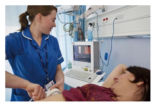
Figure 1 Transient elastography.
A specialist nurse places the probe perpendicular to the surface of the liver. A low frequency shear wave is generated along the same axis as the ultrasound transducer. The velocity of the shear wave through the liver is measured by a high frequency ultrasound signal and the output displayed as stiffness, in kPa, alongside a two-dimensional “elastogram”. The output is the median of 10 measurements, with a success rate of > 66% and an interquartile range of measurements < 1/3 of the median considered satisfactory.
- Citation: Trovato FM, Tognarelli JM, Crossey MM, Catalano D, Taylor-Robinson SD, Trovato GM. Challenges of liver cancer: Future emerging tools in imaging and urinary biomarkers. World J Hepatol 2015; 7(26): 2664-2675
- URL: https://www.wjgnet.com/1948-5182/full/v7/i26/2664.htm
- DOI: https://dx.doi.org/10.4254/wjh.v7.i26.2664


