Copyright
©The Author(s) 2015.
World J Hepatol. Nov 8, 2015; 7(25): 2578-2589
Published online Nov 8, 2015. doi: 10.4254/wjh.v7.i25.2578
Published online Nov 8, 2015. doi: 10.4254/wjh.v7.i25.2578
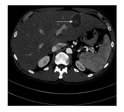
Figure 1 Contrast-enhanced computed tomography scan shows, in the region surrounding what was the probe active tip position during the ablation (white arrow), an inner hyper-dense core contrasting with an outer thicker and hypo-dense annulus.
- Citation: Poggi G, Tosoratti N, Montagna B, Picchi C. Microwave ablation of hepatocellular carcinoma. World J Hepatol 2015; 7(25): 2578-2589
- URL: https://www.wjgnet.com/1948-5182/full/v7/i25/2578.htm
- DOI: https://dx.doi.org/10.4254/wjh.v7.i25.2578


