Copyright
©The Author(s) 2015.
World J Hepatol. Aug 18, 2015; 7(17): 2069-2079
Published online Aug 18, 2015. doi: 10.4254/wjh.v7.i17.2069
Published online Aug 18, 2015. doi: 10.4254/wjh.v7.i17.2069
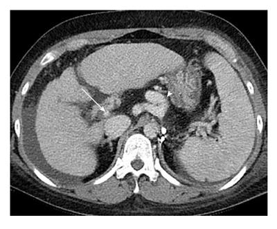
Figure 6 Image of liver cirrhosis caused by chronic hepatitis B.
Contrast enhanced computed tomography portal phase image shows the liver with irregular surface and heterogeneous enhancement of parenchyma with reticular pattern suggesting confluent fibrosis. The image shows decreased diameter of portal vein (arrow) due to large collateral vessels (arrow head) and also shows large amount of ascites.
- Citation: Yeom SK, Lee CH, Cha SH, Park CM. Prediction of liver cirrhosis, using diagnostic imaging tools. World J Hepatol 2015; 7(17): 2069-2079
- URL: https://www.wjgnet.com/1948-5182/full/v7/i17/2069.htm
- DOI: https://dx.doi.org/10.4254/wjh.v7.i17.2069


