Copyright
©The Author(s) 2015.
World J Hepatol. Aug 18, 2015; 7(17): 2069-2079
Published online Aug 18, 2015. doi: 10.4254/wjh.v7.i17.2069
Published online Aug 18, 2015. doi: 10.4254/wjh.v7.i17.2069
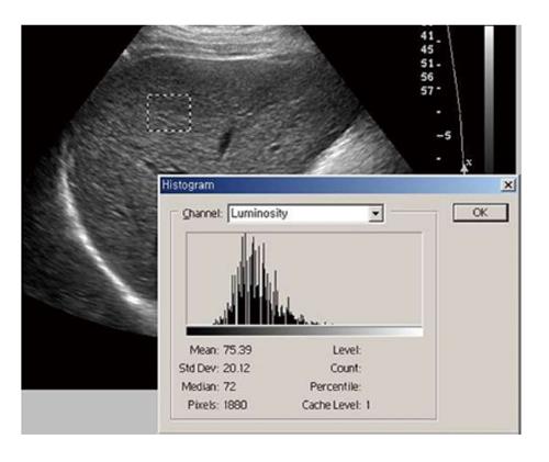
Figure 4 The region of interest of texture analysis is positioned in the right lobe of the liver, with an intercostals scan performed with gray scale ultrasonography.
Chronic liver disease patient shows heterogeneous parenchymal echogenecity with high standard deviation value (Area: 1880 pixels, Mean: 75.39, SD: 20.12).
- Citation: Yeom SK, Lee CH, Cha SH, Park CM. Prediction of liver cirrhosis, using diagnostic imaging tools. World J Hepatol 2015; 7(17): 2069-2079
- URL: https://www.wjgnet.com/1948-5182/full/v7/i17/2069.htm
- DOI: https://dx.doi.org/10.4254/wjh.v7.i17.2069


