Copyright
©The Author(s) 2015.
World J Hepatol. Aug 18, 2015; 7(17): 2069-2079
Published online Aug 18, 2015. doi: 10.4254/wjh.v7.i17.2069
Published online Aug 18, 2015. doi: 10.4254/wjh.v7.i17.2069
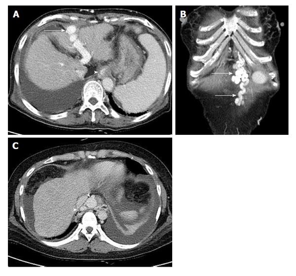
Figure 2 Image of liver cirrhosis caused by chronic hepatitis B.
Contrast enhanced computed tomography portal phase images show multiple collateral vessels of portal vein. A: The image presents large intrahepatic portosystemic shunt through left portal vein and recanalized paraumbilical vein (arrow). Lower esophageal varix is seen (arrow head); B: Coronal image shows prominent paraumbilical veins (arrows); C: Axial image shows engorged paraesophageal varix (arrow heads) which usually supplied by left gastric vein and drained into azygos- or hemiazygos-vein.
- Citation: Yeom SK, Lee CH, Cha SH, Park CM. Prediction of liver cirrhosis, using diagnostic imaging tools. World J Hepatol 2015; 7(17): 2069-2079
- URL: https://www.wjgnet.com/1948-5182/full/v7/i17/2069.htm
- DOI: https://dx.doi.org/10.4254/wjh.v7.i17.2069


